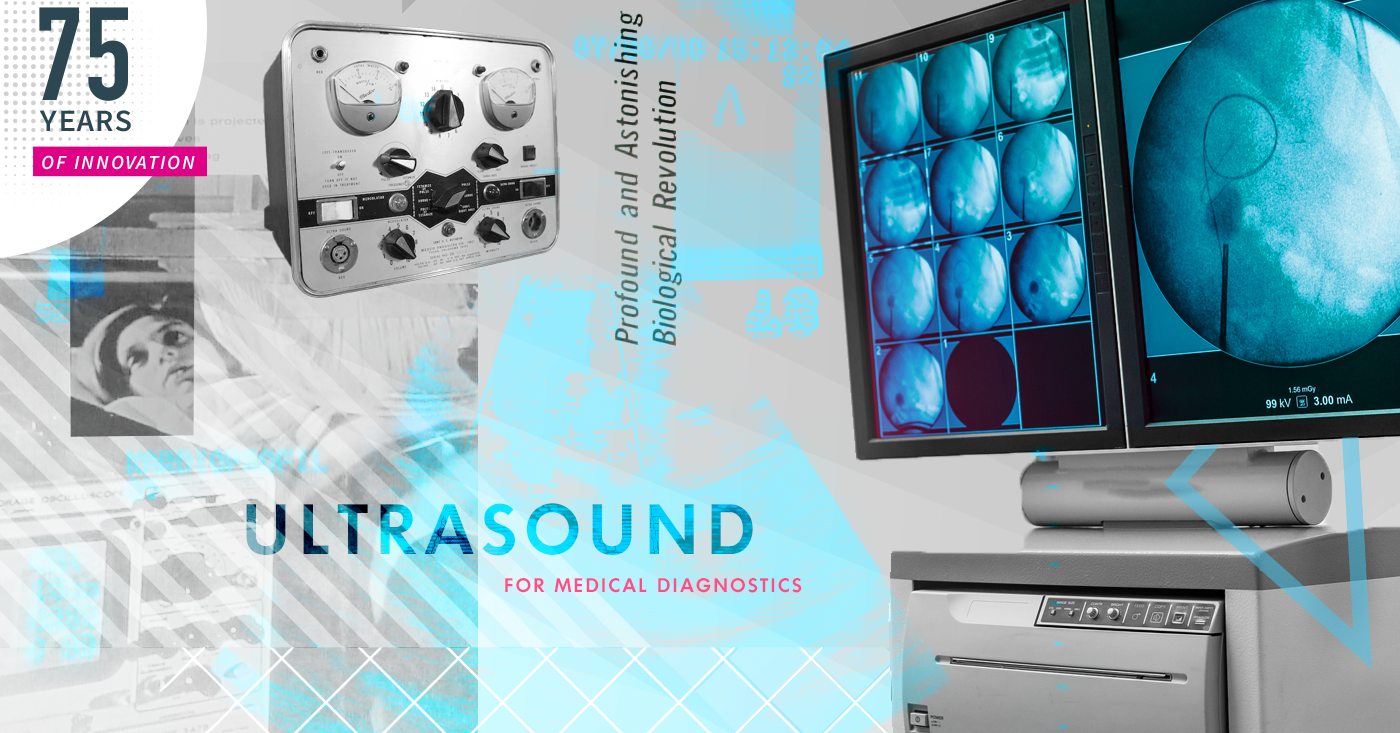
Ultrasound: A view from within
The 75 Years of Innovation series highlights the groundbreaking innovations spanning from SRI’s founding in 1946 to today. Each week, SRI will release an innovation, leading up to its 75th anniversary in November 2021.
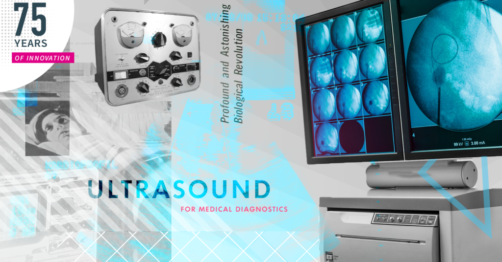
“Ultrasound is the visual stethoscope of the 21st century” — Scandinavian Journal of Trauma, Resuscitation and Emergency Medicine
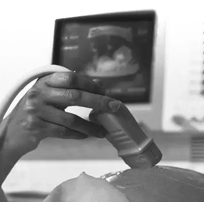
One of the most surreal and poignant moments of a pregnancy is being able to see your baby on a screen and watch as its heart takes a regular beat. This was made possible because of the medical application of a technique: ultrasound.
Ultrasound is a non-invasive way to look inside ourselves. Its application in medicine was made more effective by an amazing invention from SRI International, “Reflex Transmission Imaging” (RTI). RTI added the level of fine-grained imaging needed to make ultrasound a practical option as a ubiquitous medical diagnostic tool.
Ultrasound is used across a wide variety of medical imaging needs. It not only shows us life developing but saves lives too.
Here, we look at how ultrasound came about and what important part SRI played in making this technology an applied tool in medicine, the world over.
How Sound Becomes Images
Ultrasound is a revolution in diagnostics. It is a technique that does not require any invasive procedure to look inside a body to provide doctors with the data needed for a diagnosis. Having a way to create images of internal organs and other structures, is like switching the light on in a darkened room to find something.
The image output from an ultrasound scan is known as a ‘sonogram’. This image is created using high-frequency sound waves produced by a probe called a transducer. Typically, a transducer is placed on the skin of the patient, but for some diagnoses, to optimize the image quality, a probe can be placed inside the body, for example within the gastrointestinal tract.
A transducer contains piezoelectric crystals. When an alternating electric field is applied, these crystals vibrate, producing high-frequency sound waves. The high-frequency sound waves are directed into the body. When the sound waves hit soft tissues and other structures inside the body, an ultrasound echo is returned. This echo is, on return, detected by the piezoelectric crystals in the transducer.
Different bodily structures, such as water, bone, and soft tissue, affect the returned sound wave echo in different ways; the images are based on the speed of sound and the time taken for an echo to return. In other words, the waves that hit the transducer cause the piezoelectric crystals to create an electrical current that reflects the anatomic position and strength of the returning echoes. This is sent to the ultrasound machine which uses them to generate 2D images.
A modern transducer has a very large array of piezoelectric crystals that are activated more than 20x per second; the image can show changes over time, such as the beating heart of a fetus.
Modern ultrasound can be used for both anatomical and functional diagnosis:
- Anatomical ultrasound creates images of internal organs or other structures.
- Functional ultrasound, facilitated by RTI, is used to map things such as blood flow, tissue motion, etc. to allow physicians to visualize internal functions.
Conventional transmission imaging requires acoustic coupling to large areas on both sides of the body. Reflex Transmission Imaging (RTI) made it possible to use a single transducer for both ultrasound wave reflection and the echo return for the same region of examination, using a single transducer probe.
An Ultrasound History and SRI International
Like many things in this world, ultrasound is a product of ‘necessity being the mother of invention’. The discipline of ultrasonography is accredited to the Italian biologist Spallanzani who worked on echolocation in bats back in 1794. However, the first medical application of ultrasound was in 1942; neurologist Karl Dussik used ultrasonic waves in an attempt to detect brain tumors. Work to improve ultrasound scanners and to explore the medical role of ultrasound in areas such as midwifery, continued through the 50s and 60s. In 1958, Ian Donald with help from a commercial engineering firm, developed an automatic scanner which was used to change a clinical diagnosis of terminal cancer to a simple ovarian cyst. In 1960, the ultrasound scanner was used to make the first ante-partum diagnosis of placenta previa.
Ultrasound made major progress into the 70s and 80s, but it was the refinement of the process using Reflex Transmission Imaging that took it to new levels of diagnostic accuracy. In 1984, Philip Green and his colleagues at SRI International, refined ultrasound. They used RTI to add the capability to generate high-resolution imaging, able to show, for example, atherosclerotic plaque in arteries and the associated blood flow. Green invented this new technique, termed, “Reflex Transmission Imaging.” RTI facilitates the use of a single transducer to provide both reflective and transmissive ultrasound waves when used in the same region of examination.
RTI is used for many varied diagnoses; for example, when combined with white light digital photography, it can be used to classify pigmented lesions and detect skin melanomas.
Ultrasound is used daily throughout the world for a multitude of non-invasive diagnostic requirements. The World Health Organization (WHO) believes that ultrasound, X-ray or a combination of both, can meet two-thirds of the imaging needs of developing countries too.
The modern high-resolution ultrasound is here because of a team at SRI International. The diagnoses that ultrasound machines and the technologists and physicians that use them make, have saved many lives, and watched as many more come into the world, safely.
Resources
Gillman, L.M., Kirkpatrick, A.W. Portable bedside ultrasound: the visual stethoscope of the 21st century. Scand J Trauma Resusc Emerg Med 20, 18 (2012). https://doi.org/10.1186/1757-7241-20-18
National Institutes of Health, Philip Green, Ultrasonic Reflex Transmission Imaging, 1985: https://grantome.com/grant/NIH/R01-CA041579-01
Green, P., et.al., Combined reflection and transmission ultrasound imaging, Ultrasound in medicine and biology, 1991: https://doi.org/10.1016/0301-5629(91)90049-3
World Health Organization (WHO): https://www.who.int/medical_devices/global_forum/I03.pdf?ua=1
NIBIB, How Ultrasound Works video: https://www.youtube.com/watch?v=I1Bdp2tMFsY
Karl Theo Dussik: http://www.ob-ultrasound.net/dussikbio.html
Donald I, MacVicar J, Brown TG. Investigation of abdominal masses by pulsed ultrasound. Lancet 1958;i:1188–95.