
The 75 Years of Innovation series highlights the groundbreaking innovations spanning from SRI’s founding in 1946 to today. Each week, SRI will release an innovation, leading up to its 75th anniversary in November 2021.
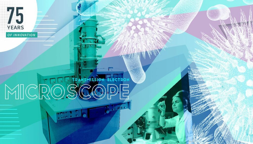
The commercialized high-resolution transmission electron microscope (TEM)
The microscope is something many of us first encounter in elementary school. You look down a short barrel into a world of strange, sometimes moving, objects. Take a drop of pond water and place it under a microscope and you see that it is filled with life. The microscope has been a vital piece of scientific equipment since its invention by Hans and Zacharias Janssen in 1590.
Over the years the microscope has evolved. Today, commercial high-performance microscopes, such as the modern Transmission Electron Microscope (TEM), are a far cry from the equipment used in classrooms. The high-resolution TEM was a product of RCA Laboratories (eventually renamed Sarnoff Corporation and now part of SRI International) back in 1940.
Today, the Transmission Electron Microscope is used for many applications including material science, medicine, biological research, forensic analysis, geology, and many other areas.
Let’s turn the lens towards the development of this important technology.
Looking down the lens into the world of the invisible
Examining the world of the tiny has challenged scientists for many centuries. An optical microscope, of the type we use in schools, uses a lens to focus light on smaller and smaller objects, eventually reaching a brick wall. The optimal resolution of an optical microscope is 0.2 microns or magnification of around 1000–2000 times. Development in the field of microscopy turned to the use of charged particles instead of light to resolve fine details in tiny specimens.
The wavelength of light limits magnification. That is, if the wavelength is larger than the tiny thing you are looking at, that thing cannot be resolved. In Transmission Electron Microscopes, light is replaced by electrons. The magnetic wavelength of an electron is extremely small, much smaller than light, and the result is observation of significantly smaller objects. A Transmission Electron Microscope (TEM) can offer resolutions of between 50 to 50 million times.
Specimen preparation for analysis using TEM is more complex than optical microscopy. The sample must be thinly sliced using an instrument such as an ultramicrotome to allow effective interaction with the electron beam. If the substance is biological, it must be preserved and dehydrated. The sample is then prepared by mounting and staining.
The TEM has four components:
- An electron source that is typically fitted to the top of the microscope.
- A vacuum tube that emits the electrons.
- The electronic components that generate the electrons and control the elector beam; the electrons being targeted using an electromagnetic lens onto the sample.
- Computer software and imaging equipment to display the specimen image in 2D (3D images can be generated by using tomographic techniques to build up a picture).
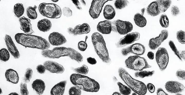
The output by a TEM is a high-resolution black and white image. The speed of the electrons in the beam correlates to wavelength. The faster an electron speed, the shorter the wavelength. Shorter wavelengths deliver more detailed images.
The electrons that pass through the specimen are captured by a detector. These “unscattered” electrons create a “shadow image”. The resulting image has light and dark areas. The light areas are where electrons have passed through the specimen, the darker areas are denser areas that prevent electron passage. The differing shades are used to determine the features of the substance.
Transmission Electron Microscopy is a developing field and advances (such as spherical aberration correction) can fine-tune resolved images and remove blurring.
The place of the Transmission Electron Microscope in history
The electron microscope is a product of the 1930s. Its history is not about time but about resolution. The goal was to be able to see the into the world of the tiny with more detail. However, the resolution of the light microscope was limited by the wavelength of light. This was resolved by turning to the very short wavelengths of an electron. The ‘focus’ in the world of microscopy has been to resolve down to atomic levels. In many ways, the use of an atomic particle, i.e., an electron, was an obvious choice in resolving an atom. However, it took many decades of experimentation to get to that point.
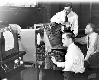
The first electron microscope was created in 1931 by two German scientists, Ernst Ruska and Max Knoll. This first attempt was not useful for any commercial endeavor, the resolution being poor, and the specimen image blurred. But it was still revolutionary in concept. Further work to improve resolution limits continued throughout the 1930s. In 1940, RCA Laboratories (later merged into SRI) commercialized the first high-resolution transmission electron microscope in North America; this TEM became known as ‘Model B’ and created a usable electron microscope for wider application.
Model B was a behemoth, measuring 10 feet high and weighing in at half a ton. It was demonstrated in Philadelphia on April 14, 1940. The RCA Transmission Electron Microscope was used successfully to observe cancer cells as early as 1949.
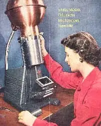
Around 2000 of RCA electron microscopes were sold before production ended in the 1960s.
The Transmission Electron Microscope continues to be used throughout a variety of areas including hospitals and research institutions.
Using a microscope may mean you look at the small, but it opens up a whole new world. Without the development of Electron Transmission Microscopes, we would be blind to many of the wonders that lie at the atomic level.
Resources
Obituary of Dr. James Hiller, New York Times: https://www.nytimes.com/2007/01/22/science/22hillier.html
RCA demonstrates electron microscope, April 14, 1940: https://www.edn.com/rca-demonstrates-electron-microscope-april-14-1940/
E J. Big., A Short History of the Electron Microscope, Bios, vol. 27, no. 1, 1956, pp. 33–37: JSTOR, www.jstor.org/stable/4605737


